42 results filtered with: Connections
- Digital Images
- Online
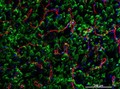
Cellular architecture of normal human skin imaged by whole mount tissue microscopy. Human skin has a rich network of white blood cells (specifically dendritic cells, T cells and macrophages) which form sheaths around blood vessels. This image was taken directly beneath the junction that joins the dermal and epidermal layers of the skin (dermo-epidermal junction). At this level, the capillary network (stained for CD31; red) is visualised against a lawn of autofluorescent dermal papillae (finger-like projections of the dermis; green) scattered with dendritic cells (stained for CD11c; green) and macrophages (stained for LYVE-1; blue). This normal cellular architecture is grossly disrupted in diseased skin (see related images). Scale bar (white) represents 200 micrometres.
Dr. Xiao-nong Wang, Human Dendritic Cell Laboratory, Newcastle University
- Digital Images
- Online
Arcuate Fasiculus, tractography
Katja Heuer and Roberto Toro
- Digital Images
- Online
Cellular architecture of normal human skin imaged by whole mount tissue microscopy. Human skin has a rich network of white blood cells (specifically dendritic cells, T cells and macrophages) which form sheaths around blood vessels. In this image, blood vessels (string-like structures stained for CD31; green), lymphatic vessels (ribbon-like structures stained for LYVE-1; blue) and T cells (stained for CD3; red) can be seen. T cells are only found around dermal blood vessels. Macrophages (stained for LYVE-1; blue) are also present. This normal cellular architecture is grossly disrupted in diseased skin (see related images). X10 magnification. Scale bar (white) represents 200 micrometres.
Dr. Xiao-nong Wang, Human Dendritic Cell Laboratory, Newcastle University
- Digital Images
- Online
Corticospinal tract, tractography
Katja Heuer and Roberto Toro
- Digital Images
- Online
Corpus Callosum, tractography
Katja Heuer and Roberto Toro
- Digital Images
- Online
Synaptic connection in rat cerebellar cortex
Prof. M. Hausser / UCL
- Digital Images
- Online
Unfolded brain, MRI
Katja Heuer and Roberto Toro
- Digital Images
- Online
Neuronal migration is an artwork depicting many very young neurons that have been produced in the neuroepithelium migrating to their appropriate destinations in the brain. This image highlights the future of neuroscience showing different classes of cells colour coded. There is no available technique to do this now, but it is not far off considering the advances that have been made with brainbow mice. The brainbow technique allows for different cell types to be tagged with fluorescent proteins to track their development and connections with other cells.
Prof. Bill Harris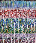
- Digital Images
- Online
Mueller glial cells in the retina
Prof. Bill Harris
- Digital Images
- Online
Cell fates in zebrafish retina, acrylic painting
Prof. Bill Harris
- Digital Images
- Online
Insomnia
Stephen Magrath
- Digital Images
- Online
White matter innervation of the neocortex, MRI
Katja Heuer and Roberto Toro
- Digital Images
- Online
Pyramidal neuron
Prof. M. Hausser / UCL
- Digital Images
- Online
Cellular architecture of normal human skin imaged by whole mount tissue microscopy. Human skin has a rich network of white blood cells (specifically dendritic cells, T cells and macrophages) which form sheaths around blood vessels. In this image, blood vessels (string-like structures stained for CD31; red), lymphatic vessels (ribbon-like structures stained for LYVE-1; blue) and dendritic cells (stained for CD11c; green) can be seen. Macrophages (stained for LYVE-1; blue) are also present. This normal cellular architecture is grossly disrupted in diseased skin (see related images). X10 magnification. Scale bar (white) represents 200 micrometres.
Dr. Xiao-nong Wang, Human Dendritic Cell Laboratory, Newcastle University
- Digital Images
- Online
Neurone development, embryoid body
John Grady, Doug Turnbull, Claudia Racca, Newcastle University
- Digital Images
- Online
Cellular architecture of normal human skin imaged by whole mount tissue microscopy. Human skin has a rich network of white blood cells (specifically dendritic cells, T cells and macrophages) which form sheaths around blood vessels. This image was taken greater than 150 micrometres beneath the junction that joins the dermal and epidermal layers of the skin (dermo-epidermal junction). At this level, dendritic cells (stained for CD11c; green) and macrophages (stained for LYVE-1; blue) form clusters around blood vessels (stained for CD31; red). This normal cellular architecture is grossly disrupted in diseased skin (see related images). Scale bar (white) represents 100 micrometres.
Dr. Xiao-nong Wang, Human Dendritic Cell Laboratory, Newcastle University
- Digital Images
- Online
Silicon chip
Paul Griggs